The first morning specimen of urine is more
concentrated and will have a higher specific gravity
than a specimen passed during the day. A high fluid
intake may reduce the specific gravity to below 1.010.
In the presence of disease, the specific gravity of a
24-hour specimen may vary from 1.001 to 1.060.
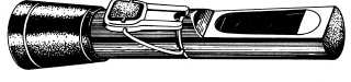
Specific gravity is measured with an index
refractometer, available as standard equipment at most
duty stations. See figure 7-22. The index refracto-
meter may be held manually or mounted on a stand like
a microscope. The specific gravity of urine is
determined by the index of light refraction through
solid material.
Measure the specific gravity with an index
refractometer in the following manner:
1. Hold the index refractometer in one hand. Use
the other hand and an applicator stick to place a
drop of urine on the glass section beneath the
coverglass.
2. Hold the refractometer so that the light reflects
on the glass section, and look into the ocular
end. Read the number that appears where the
light and dark lines meet. This is the specific
gravity.
Chemical Characteristics
Chemical characteristics evaluated during a
routine urinalysis include pH, protein, glucose,
ketones, and blood. Some laboratories also include
tests for bilirubin, urobilinogen, and nitrite, depending
on the test strip used.
Currently, most medical
facilities use the Multistix® and Color Chart, which
detects pH, protein, glucose, ketones, blood, bilirubin,
and urobilinogen.
The Multistix is a specially
prepared multitest strip. The strip is simply dipped
into the urine specimen and compared to the color
values for the various tests on the accompanying chart.
The color chart also indicates numerical pH values,
which should be reported.
Microscopic Examination of Urine Sediment
Microscopic examination of urine sediment is
usually performed in addition to routine procedures.
This examination requires a degree of skill acquired
through practice under the immediate supervision of
an experienced technician. The specimen used for
microscopic examination should be as fresh as
possible. Red cells and many formed solids tend to
disintegrate upon standing, particularly if the
specimen is warm or alkaline.
PREPARING SPECIMENS FOR MICRO-
SCOPIC EXAMINATION.—To prepare urine
specimens for microscopic examination, follow the
steps below.
1. Stir the specimen well.
2. Pour 15 ml of urine into a conical centrifuge
tube, and centrifuge at 1,500 rpm for 5 minutes.
3. Invert the centrifuge tube and allow all of the
excess urine to drain out. Do not shake the tube
while it is inverted. Enough urine will remain
in the tube to resuspend the sediment. Too much
urine will cause dilution of the sediment,
making an accurate reading difficult.
4. Resuspend the sediment by tapping the bottom
of the tube.
5. With a medicine dropper, mount one drop of the
suspension on a slide and cover it with a
coverslip.
6. Place the slide under the microscope, and scan
with the low-power objective and subdued
lighting.
7. Switch to the high-power objective for detailed
examination of a minimum of 10 to 15 fields.
CLINICALLY SIGNIFICANT FINDINGS.—
Leukocytes, erythrocytes, and casts may all be of
clinical significance when found in urine sediment.
Leukocytes.—Normally, 0 to 3 leukocytes per
high-power field will be seen on microscopic
examination. More than 3 cells per high-power field
probably indicates disease somewhere in the urinary
tract. Estimate the number of leukocytes present per
high-power field and report it as the “estimated
number per high-power field.”
Erythrocytes.—Red cells are not usually present
in normal urine. If erythrocytes are found, estimate
their number per high-power field and report it.
Erythrocytes may be differentiated from white cells in
several ways:
White cells are larger than red cells.
7-35
HM3f0722
Figure 7-22.—Index refractometer.