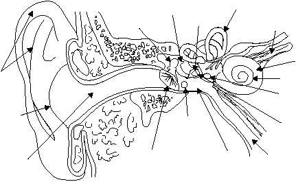
(fig. 1-52). The malleus is attached to the inner surface
of the eardrum and connects with the incus, which in
turn connects with the stapes. The base of the stapes is
attached to the fenestra ovalis (oval window), the
membrane-covered opening of the inner ear. These
tiny bones, which span the middle ear, are suspended
from bony walls by ligaments. This arrangement
provides the mechanical means for transmitting sound
vibrations to the inner ear.
The eustachian tube, or auditory tube, connects
the middle ear with the pharynx. It is lined with a
mucous membrane and is about 36 mm long. Its
function is to equalize internal and external air
pressure. For example, while riding an elevator in a tall
building, you may experience a feeling of pressure in
the ear. This condition is usually relieved by
swallowing, which opens the eustachian tube and
allows the pressurized air to escape and equalize with
the area of lower pressure. Divers who ascend too fast
to allow pressure to adjust may experience rupture of
their eardrums. The eustachian tube can also provide a
pathway for infection of the middle ear.
Inner Ear
The inner ear is filled with a fluid called
endolymph. Sound vibrations that cause the stapes to
move against the oval window create internal ripples
that run through the endolymph. These pressurized
ripples move to the cochlea, a small snail-shaped
structure housing the organ of Corti, the hearing
organ (fig. 1-52). The cells protruding from the organ
of Corti are stimulated by the ripples to convert these
mechanical vibrations into nerve impulses, and these
impulses are relayed through the vestibulocochlear
(8th cranial) nerve to the auditory area of the cortex in
the temporal lobe of the brain. There they are
interpreted as the sounds we hear.
Another structure located in the inner ear is
composed of the three semicircular canals, situated
perpendicular to each other. Movement of the
endolymph within the canals, caused by general body
movements, stimulates nerve endings, which report
these changes in body position to the brain, which in
turn uses the information to maintain equilibrium.
The fenestra rotunda (round window) is another
membrane-covered opening of the inner ear. It
contracts the middle ear and flexes to accommodate
the inner ear ripples caused by the stapes.
TOUCH
Until the beginning of the last century, touch
(feeling) was treated as a single sense. Thus, warmth or
coldness, pressure, and pain, were thought to be part of
a single sense of touch or feeling. It was discovered
that different types of nerve ending receptors are
widely and unevenly distributed in the skin and
mucous membranes. For example, the skin of the back
possesses relatively few touch and pressure receptors
while the fingertips have many. The skin of the face has
relatively few cold receptors, and mucous membranes
have few heat receptors. The cornea of the eye is
sensitive to pain, and when pain sensation is abolished
by a local anesthetic, a sensation of touch can be
experienced.
1-48
HM3F0152
MALLEUS
INCUS
STAPES
SEMICIRCULAR
CANALS
INNER
EAR
VESTIBULOCOCHLEAR
NERVE
COCHLEA
OVAL
WINDOW
ROUND
WINDOW
TYMPANIC
CAVITY
MIDDLE
EAR
TYMPANIC
MEMBRANE
EXTERNAL
AUDITORY
CANAL
OUTER
EAR
AURICLE
PHARYNX
Figure 1-52.—Major parts of the ear.