THE SENSORY SYSTEM
LEARNING OBJECTIVE: Recognize the
senses of the body, and identify their physical
characteristics.
The sensory system informs areas of the cerebral
cortex of changes that are taking place within the body
or in the external environment. The special sensory
receptors respond to special individual stimuli such as
sound waves, light, taste, smell, pressure, heat, cold,
pain, or touch. Positional changes, balance, hunger,
and thirst sensations are also detected and passed on to
the brain.
SMELL
Odor is perceived upon stimulation of the receptor
cells in the olfactory membrane of the nose. The
olfactory receptors are very sensitive, but they are
easily fatigued. This tendency explains why odors that
are initially very noticeable are not sensed after a short
time. Smell is not as well developed in man as it is in
other mammals.
TASTE
The taste buds are located in the tongue. The
sensation of taste is limited to sour, sweet, bitter, and
salty. Many foods and drinks tasted are actually
smelled, and their taste depends upon their odor. (This
interdependence between taste and smell can be
demonstrated by pinching the nose shut when eating
onions.) Sight can also affect taste. Several drops of
green food coloring in a glass of milk will make it all
but unpalatable, even though the true taste has not been
affected.
SIGHT
The eye, the organ of sight, is a specialized
structure for the reception of light. It is assisted in its
function by accessory structures, such as the eye
brows, eyelashes, eyelids, and lacrimal apparatus.
The lacrimal apparatus consists of structures that
produce tears and drains them from the surface of the
eyeball.
Structure of the Eye
Approximately five-sixths of the eyeball lies
recessed in the orbit, protected by a bony socket. Only
the small anterior surface of the eyeball is exposed.
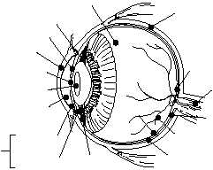
The eye is not a solid sphere but contains a large
interior cavity that is divided into two cavities, anterior
and posterior. The anterior cavity is further subdivided
into anterior and posterior chambers (fig. 1-48).
The anterior cavity of the eye lies in front of the
lens. The anterior chamber of the anterior cavity is
the space anterior to the iris, but posterior to the cornea.
The posterior chamber of the anterior cavity consists
of a small space directly posterior to the iris, but
anterior to the lens. Both chambers of the anterior
cavity are filled with a clear, watery fluid called
aqueous humor. Aqueous humor helps to give the
cornea its curved shape.
The posterior cavity of the eye is larger than the
anterior cavity, since it occupies all the space posterior
to the lens, suspensory ligaments, and ciliary body. The
posterior cavity contains a substance, with the
consistency similar to soft gelatin, called vitreous
humor. Vitreous humor helps maintain sufficient
pressure inside the eye to prevent the eyeball from
collapsing.
The eyeball is composed of three layers. From the
outside in, they are the sclera, choroid, and retina (fig.
1-48).
OUTER LAYER.—The outer layer of the eye is
called the sclera. The sclera is the tough, fibrous,
protective portion of the globe, commonly called the
white of the eye. Anteriorly, the outer layer is
transparent and is called the cornea, or the window of
the eye. It permits light to enter the globe. The exposed
sclera is covered with a mucous membrane, the
conjunctiva, which is a continuation of the inner lining
of the eyelids. The lacrimal gland produces tears that
constantly wash the front part of the eye and the
conjunctiva. The tear gland secretions that do not
1-45
HM3F0148
VITREOUS
HUMOR
SCLERA
OPTIC
DISK
OPTIC
NERVE
FOVEA
CENTRALIS
POSTERIOR
CAVITY
RETINA
CHOROID
COAT
CILIARY
BODY
ANTERIOR
CHAMBER
POSTERIOR
CHAMBER
ANTERIOR
CAVITY
AQUEOUS
HUMOR
LENS
PUPIL
IRIS
CORNEA
SUSPENSORY
LIGAMENTS
Figure 1-48.—Transverse section of the eye.