THE MICROSCOPE
LEARNING OBJECTIVE:
Identify the
parts of the microscope, and determine their
functions.
Before any attempts are made to view blood
smears, urinary sediments, bacteria, parasites, etc., it is
absolutely essential that beginners know the
instrument with which they will be spending
considerable time—the microscope. The microscope
is a precision instrument used extensively in clinical
laboratories to make visible objects too small to be
seen by the unaided eye. Most laboratories are
equipped with binocular (two-eyepiece) microscopes,
but monocular microscopes are also commonly used.
The type of microscope most often used in the
laboratory is referred to as the compound microscope.
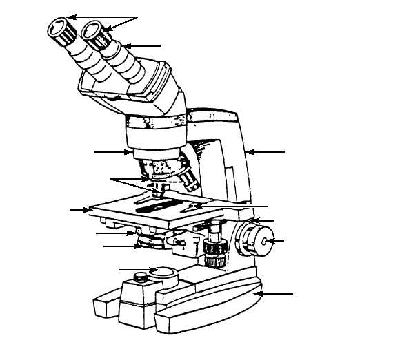
See figure 7-5. A compound microscope contains a
system of lenses of sufficient magnification and
resolving power (ability to show, separate, and
distinguish) so that small elements lying close together
in a specimen appear larger and distinctly separated. In
the following sections, the compound microscope’s
framework, illumination system, magnification
system, and focusing system will be discussed.
FRAMEWORK
The framework of the compound microscope
consists of four parts:
the arm, the stage, the
mechanical stage, and the base (fig. 7-5).
Arm
The arm is the structure that supports the
magnification and focusing system. It is the handle by
which the microscope is carried.
Stage
The stage is the platform on which a specimen is
placed for examination. In the center of the stage is an
aperture or hole that allows the passage of light from
the condenser.
7-7
EYEPIECES
BODY TUBE
ARM
MECHANICAL STAGE
COARSE CONTROL KNOB
FINE CONTROL KNOB
BASE
INTERNAL
LIGHT SOURCE
CONDENSER
IRIS DIAPHRAGM
STAGE
OBJECTIVES
REVOLVING
NOSEPIECE
HM3f0705
Figure 7-5.—Compound microscope.