you are making the smear, prevent blood from
reaching the extreme edges of the slides.
Allowing the smear to reach the edges of the
slide will aggravate the tendency of large cells to
stack up on the perimeter of the smear. A smear
with wavy lines or blanks spots should be
discarded, and a new smear made.
5. Once the blood smear is made, let it dry (it will
take a few minutes). Then write the patient’s
name in pencil on the bottom edge of the slide,
as illustrated in figure 7-18, view D). Proceed to
step 2, staining the cells.
STAINING THE CELLS.—Once a blood smear
is made, it should be stained. Staining the blood smear
highlights the differences among the different types of
leukocytes for easier recognition during the counting
process. The most popular stain used for this purpose
is Wright’s stain. Wright’s stain is a methyl alcohol
(methanol) solution of an acid dye and a basic dye. The
acid dye in Wright’s stain is known as eosin and is red
in color. The basic dye in Wright’s stain is known as
methylene blue and is blue in color. Generally, white
cells are identified by their affinity to the dye they
prefer. For example, cells that prefer the acid dye
(eosin) are called eosinophils. Other cells that prefer
the basic dye are called basophils.
WARNING
Wright’s staining solution contains methanol, which
is considered a hazardous material. It is classified as
flammable, a poison, and an irritant. Methanol must
be kept away from heat, sparks, and open flames.
Good ventilation in usage areas is paramount since
exposure to vapors can irritate eyes, nose, throat, and
mucous membranes of the upper respiratory tract.
When not in use, methanol containers should be
closed tightly and stored upright to prevent leakage.
Gloves and protective clothing (e.g., lab coat or
apron) and eyewear should be worn to avoid contact
with the solution. Absorption through skin can cause
permanent blindness. Death may result from
ingestion or exposure to high vapor concentrations of
methanol.
There are a variety of staining products on the
market today. Some of these staining products have
combined Wright’s solution with other staining
solutions, such as Giemsa stain. When using a new
product, you should always review the manufacturer’s
usage and safety recommendations.
The staining process that we will cover in this
chapter is known as a quick stain. A quick stain has
very few equipment requirements and only a few
procedural steps. An example of a quick stain is One
Step II Wright-Giemsa Stain Solution® by Criterion
Sciences. To stain a blood smear with this product,
follow the steps below.
1. Prepare two staining containers by filling one
with One Step II stain solution and the other
with deionized or distilled water. The use of tap
water instead of deionized or distilled water is
not recommended since the pH of tap water
varies.
If tap water is used, its pH should
between 5.8 and 7.03.
2. Immerse the slide (blood smear) in the stain for
15 to 30 seconds.
(To prevent debris or
precipitate from contaminating the slide, do not
add new stain to old.)
3. Remove the slide and allow excess stain to drain
from the edge of the slide.
4. Immerse the slide in the deionized or distilled
water for 5 to 15 seconds. (Change the water
7-22
A
B
C
D
HM3f0718
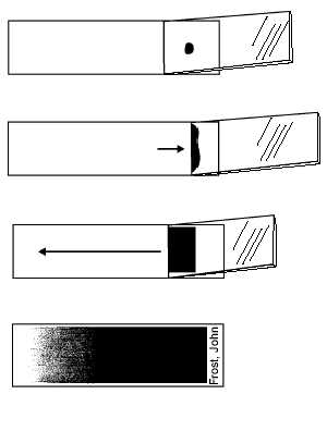
Figure 7-18.—Making a blood smear: A. Placing second slide
at a 23Eangle; B. Blood distributing itself along second slide’s
edge; C. Drawing blood across surface of slide; D. Example
of a properly prepared blood smear.