when it becomes dark blue or when film forms
on the surface.) NOTE: Rinse time is critical
and must be shorter than the stain time.
5. Drain excess water and wipe the back of the
slide to reduce background color.
6. Place slide in horizontal position on table and
allow to air dry.
NOTE: Do not accelerate
drying time by placing slide on a warmer or in
front of a fan. The film of water on the slide is
important for the color development.
7. Once the slide is dry, proceed to step 3, counting
the cells.
COUNTING THE CELLS.—Once the blood
smear has been stained, it is placed under a
microscope, and the differential count is conducted.
To perform a differential white cell count, you
should follow the steps listed below:
1. Place the slide under the microscope. Switch
the oil immersion objective (red) (100X) into
position above the stage. Turn the coarse
adjustment to raise the oil immersion objective
about 1 inch above the opening in the stage.
Open the condenser and switch on the
microscope light.
2. Place a large drop of immersion oil on the thin
area of the blood smear. See figure 7-19.
3. Hold the slide so the thin area is on your left.
Then fix the slide firmly in the jaws of the
mechanical (movable) stage.
Move the
mechanical stage so the drop of oil on the slide is
directly over the bright light coming up from the
condenser.
4. Using the coarse control knob, you should now
slowly lower oil immersion objective into the
drop of oil (on the slide). When the objective is
in the drop of oil, continue turning the coarse
adjustment until the objective is touching the
glass slide.
5. Now, while continually looking through the
eyepiece, VERY SLOWLY rotate the coarse
adjustment toward you until you see some cells.
After you have brought the cells into view with
the coarse adjustment, bring the cells into
perfect focus by rotating the fine adjustment.
NOTE: Always rotate the fine adjustment back
and forth when identifying cells. This step will
help you see the various layers of the cell and
thereby help you to identify the different types
of white cells.
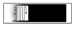
6. Count 100 consecutive white cells, pressing the
correct key on the cell counter for each type of
white cell identified. (If the cell counter is not
available, record cell type and number of cells
encountered on a piece of paper.) Follow path
similar to one illustrated in figure 7-20 to count
cells.
7. Total each type of white cell. If you count 20
lymphocytes among the 100 cells, the
differential count for lymphocytes is 20%.
Continue this process until your count totals
100%. This differential count is referred to as a
relative count. Another differential count that
may be requested is an absolute count. To
perform an absolute count, multiply the total
white cell count by the individual cell
percentages. See the example below.
7-23
DROP OF IMMERSION OIL
HM3f0719
THIS THIN AREA OF THE
BLOOD SMEAR IS FOR
IDENTIFYING THE CELLS
Figure 7-19.—Placement of immersion oil on blood smear.
HM3f0720
Figure 7-20.—Counting path for differential count.
Example:
Patient has a total white cell count of 8,000.
Differential count shows 20% leukocytes.
Multiply:
8,000 x 0.20 (20%) = 1,600
Patient has 1,600 lymphocytes/mm3