of female secondary sexual characteristics. See section
titled “Endocrine System” for listing of secondary
female sexual characteristics.
The ovaries are also the primary source of
progesterone (in a nonpregnant female). This hormone
promotes changes that occur in the uterus during the
female reproductive cycle. In addition, progesterone
stimulates the enlargement of mammary glands and
ducts, and increases fat deposits in female breasts
during puberty.
INTERNAL ACCESSORY ORGANS
The internal accessory organs of the female
reproductive system include a pair of fallopian tubes,
the uterus, and the vagina (fig. 1-61).
Fallopian Tubes
The fallopian tubes, also known as uterine tubes,
serve as ducts for the ovaries, providing a passageway
to the uterus. The fallopian tubes are composed of three
tissue layers. These tissue layers include an inner
mucosal layer, a middle muscular layer, and an outer
serous layer, and they are continuous with the layers of
the uterus. The fallopian tubes are in contact with the
ovaries but are not continuous with them. Their
funnel-shaped openings, called free openings, are
fringed with fingerlike processes that pick up an ovum
and draw it into the fallopian tubes. Once the ovum
enters the fallopian tubes, it is transported to the uterus
by peristalsis and gravity. Fertilization of an ovum
normally takes place in the fallopian tubes.
Uterus
The function of the uterus is to receive the embryo
that results from the fertilization of an egg cell, and to
sustain its life during development. The uterus, or
womb, is a hollow, pear-shaped organ with thick,
muscular walls. The uterus is divided into two main
regions, the body and cervix (fig. 1-61). The body of
the uterus consists of the upper two-thirds of the uterus.
The cervix is the lower one-third portion of the uterus
that projects into the upper part of the vagina. The
cervical opening into the vagina is called the external
os.
The uterine wall is composed of three layers: the
endometrium, the myometrium, and the perimetrium.
The inner lining consists of specialized epithelium,
called endometrium, which undergoes partial
destruction approximately every 28 days in the
n o n p r e g n a n t f e m a l e . T h e m i d d l e l a y e r, t h e
myometrium, consists of bundles of interlaced
muscular fibers. The muscular layer produces
powerful rhythmic contractions that are important in
the expulsion of the fetus at birth. The perimetrium
consists of an outer serosal layer that covers the body
of the uterus and part of the cervix. The uterus also has
three openings: superiorly and laterally, two openings
connect the fallopian tubes to the uterus, and inferiorly,
an opening leading to the vagina.
1-61
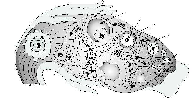
FOLLICULAR
FLUID
OOCYTE
FOLLICULAR
CELLS
OVARY
PRIMORDIAL
FOLLICLE
TIME
OVULATION
FALLOPIAN
TUBE
HM3F0162
Figure 1-62.—Ovulation process.