bones return to their normal position, forcing air from
the lungs.
THE PROCESS OF RESPIRATION
The rhythmical movements of breathing are
controlled by the respiratory center in the brain.
Nerves from the brain pass down through the neck to
the chest wall and diaphragm. The nerve that controls
the diaphragm is called the phrenic nerve; the nerve
that controls the larynx is the vagus nerve; and the
nerves that control the muscles between the ribs are the
intercostal nerves.
The respiratory center is stimulated by chemical
changes in the blood. When too much carbon dioxide
accumulates in the blood stream, causing the blood to
become acidic, the respiratory center signals the lungs
to breathe faster to get rid of the carbon dioxide.
The respiratory center can also be stimulated or
depressed by a signal from the brain. For example,
changes in one's emotional state can alter respiration
through laughter, crying, emotional shock, or panic.
The muscles of respiration normally act
automatically, with normal respiration being 14 to 18
cycles per minute. The lungs, when filled to capacity,
hold about 6,500 ml of air, but only 500 ml of air is
exchanged with each normal respiration. This
exchanged air is called tidal air. The amount of air left
in the lungs after forceful exhalation is about 1,200 ml
and is known as residual air.
THE NERVOUS SYSTEM
LEARNING OBJECTIVE: Identify the
components and function of a neuron, recall the
process of impulse transmission, and identify
the components and functions of the central
and peripheral nervous systems.
The activities of the widely diverse cells, tissues,
and organs of the body must be monitored, regulated,
and coordinated to effectively support human life. The
interaction of the nervous and endocrine systems
provides the needed control.
The nervous system is specifically adapted to the
rapid transmission of impulses from one area of the
body to another. On the other hand, the endocrine
system, working at a far slower pace, maintains body
metabolism at a fairly constant level.
In this section, you will study the neuron, the basic
functional unit of the nervous system. Also, you will
study the components and functions of the different
divisions of the nervous system. The nervous system is
divided into two major groups, the central nervous
system (CNS) and the peripheral nervous system
(PNS). Another division of the nervous system is the
autonomic nervous system (ANS), which is further
subdivided into the sympathetic and parasym-
pathetic nervous systems.
THE NEURON
The structure and functional unit of the nervous
system is the nerve cell, or neuron, which can be
classified into three types. The first is the sensory
neuron, which conveys sensory impulses inward from
the receptors. The second is the motor neuron, which
carries command impulses from a central area to the
responding muscles or organs. The third type is the
interneuron, which links the sensory neurons to the
motor neurons.
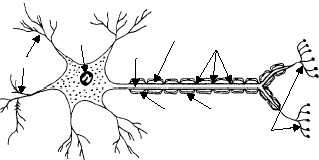
The neuron is composed of dendrites, a cyton, and
an axon (fig. 1-42). The dendrites are thin receptive
branches, and vary greatly in size, shape, and number
with different types of neurons. They serve as
receptors, conveying impulses toward the cyton. The
cyton is the cell body containing the nucleus. The
single, thin extension of the cell outward from the
cyton is called the axon. It conducts impulses away
from the cyton to its terminal branches, which
transmit the impulses to the dendrites of the next
neuron.
Large axons of the peripheral nerves are
commonly enclosed in a sheath, called neurilemma,
composed of Schwann cells (fig. 1-42). Schwann cells
wrap around the axon and act as an electrical insulator.
1-36
HM3F0142
MYELIN
SHEATH
AXON
CYTON
DENDRITES
NODES OF
RANVIER
NEURILEMMA
SCHWANN
CELL
TERMINAL
BRANCHES
Figure 1-42.—The neuron and its parts.