middle layer; and the pia mater is the vascular
inner-most layer that adheres to the surface of the brain
and spinal cord. Inflammation of the meninges is
called meningitis. The type of meningitis contracted
depends upon whether the brain, spinal cord, or both
are affected, as well as whether it is caused by viruses,
bacteria, protozoa, yeasts, or fungi.
CEREBROSPINAL FLUID.—Cerebrospinal
fluid is formed by a plexus, or network, of blood
vessels in the central ventricles of the brain. It is a clear,
watery solution similar to blood plasma. The total
quantity of spinal fluid bathing the spinal cord is about
75 ml. This fluid is constantly being produced and
reabsorbed. It circulates over the surface of the brain
and spinal cord and serves as a protective cushion as
well as a means of exchange for nutrients and waste
materials.
Spinal Cord
The spinal cord is continuous with the medulla
oblongata and extends from the foramen magnum,
through the atlas, to the lower border of the first lumbar
vertebra, where it tapers to a point. The spinal cord is
surrounded by the bony walls of the vertebral canal
(fig. 1-44). Ensheathed in the three protective
meninges and surrounded by fatty tissue and blood
vessels, the cord does not completely fill the vertebral
canal, nor does it extend the full length of it. The nerve
roots serving the lumbar and sacral regions must pass
some distance down the canal before making their exit.
The sympathetic trunk contains the paravertebral
ganglia (sing. ganglion), knotlike masses of nerve cell
bodies (fig. 1-44).
A cross section of the spinal cord shows white and
gray matter (fig. 1-45). The outer white matter is
composed of bundles of myelinated nerve fibers
arranged in functionally specialized tracts. It
establishes motor communication between the brain
and the body parts. The inner gray unmyelinated
1-38
HM3f0143
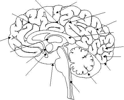
MOTOR AREAS INVOLVED WITH THE
CONTROL OF VOLUNTARY MUSCLES
CONCENTRATION, PLANNING,
PROBLEM SOLVING
FRONTAL
LOBE
MOTOR SPEECH AREA
(BROCA'S AREA)
INTERPRETATION OF SENSORY EXPERIENCES,
MEMORY OF VISUAL AND AUDITORY PATTERNS
TEMPORAL
LOBE
BRAIN
STEM
VISUAL
AREA
VISUAL IMAGES,
VISUAL RECOGNITION
OF OBJECTS
OCCIPITAL
LOBE
GENERAL
INTERPRETATIVE
AREA
PARIETAL
LOBE
UNDERSTANDING SPEECH,
USING WORDS
SENSORY AREA INVOLVED WITH
CUTANEOUS AND OTHER SENSES
CEREBELLUM
AUDITORY
AREA
Figure 1-43.—Functional areas of the brain.
HM3F0144
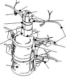
SYMPATHETIC
TRUNK
PARAVERTEBRAL
GANGLION
SPINE
NERVES
TRANSVERSE
PROCESS
INTERVERTEBRAL
FORAMEN
POSTERIOR
ROOT
DORSAL
ROOT
GANGLION
PIA
MATER
ARACHNOID
MATER
DURA
MATER
VERTEBRAL
CANAL
BODY OF
VERTEBRA
ANTERIOR
ROOT
SPINAL
CORD
Figure 1-44.—Spinal cord.