that fits into the acetabulum. Two processes called the
greater and lesser trochanters are at the proximal end
for the attachment of muscles. The neck of the femur,
located between the head and the trochanters, is the site
on the femur most frequently fractured. At the distal
end are two bony prominences, called the lateral and
medial condyles, which articulate with the tibia and
the patella.
Patella.—The patella is a small oval-shaped bone
overlying the knee joint. It is enclosed within the
tendon of the quadriceps muscle of the thigh. Bones
like the patella that develop within a tendon are known
as sesamoid bones.
Tibia.—The tibia, or shin bone, is the larger of the
two leg bones and lies at the medial side. The proximal
end articulates with the femur and the fibula. Its distal
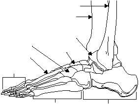
end articulates with the talus (one of the foot bones)
and the fibula (fig. 1-25). A prominence easily felt on
the inner aspect of the ankle is called the medial
malleolus.
Fibula.—The fibula, the smaller of the two leg
bones, is located on the lateral side of the leg, parallel
to the tibia. The prominence at the distal end forms the
outer ankle and is known as the lateral malleolus.
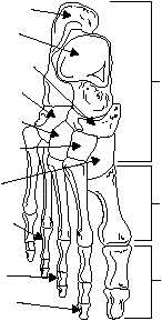
Tarsus.—The tarsus, or ankle, is formed by seven
tarsal bones: medial cuneiform, intermediate
cuneiform, lateral cuneiform, cuboid, navicular,
talus, and calcaneus. The strongest of these is the heel
bone, or calcaneus.
Metatarsus.—The sole and instep of the foot is
called the metatarsus and is made up of five
metatarsal bones (fig. 1-25). They are similar in
arrangement to the metacarpals of the hand.
Phalanges.—The phalanges are the bones of the
toes and are similar in number, structure, and
arrangement to the bones of the fingers.
JOINTS
LEARNING OBJECTIVE: Recognize joint
classifications and identify joint movements for
the key joints in the body.
Wherever two or more bones meet, a joint is
formed. A joint binds various parts of the skeletal
system together and enables body parts to move in
response to skeletal muscle contractions.
JOINT CLASSIFICATIONS
Joints are classified according to the amount of
movement they permit (fig. 1-26). Joint classifications
are as follows:
Immovable. Bones of the skull are an example
of an immovable joint. Immovable joints are
characterized by the bones being in close contact with
each other and little or no movement occurring between
the bones.
Slightly movable. In slightly movable joints,
the bones are held together by broad flattened disks of
cartilage and ligaments (e.g., vertebrae and symphysis
pubis).
1-15
HM3f0125
METATARSUS
TARSUS
PHALANGES
METATARSALS
MEDIAL
CUNEIFORM
NAVICULAR
TALUS
TIBIA
FIBULA
CALCANEUS
TALUS
NAVICULAR
CUBOID
LATERAL
CUNEIFORM
INTERMEDIATE
CUNEIFORM
MEDIAL
CUNEIFORM
PROXIMAL
PHALANX
MIDDLE
PHALANX
DISTAL
PHALANX
TARSALS
METATARSALS
PHALANGES
Figure 1-25.—The foot: A. Lateral view of foot; B. Right foot viewed from above.


