the long axis of the tooth being X-rayed. A tube head with a 16-inch X-ray source to cylinder end distance (long cone) should be used with the paralleling technique. The tube head must be positioned so that the central X-ray beam is projected perpendicular to the tooth and the film packet. To properly position the film and the tube head, use paralleling devices.
There are two different paralleling devices. One is used for radiographs of the anterior teeth; the other is used for radiographs of the posterior teeth. Each paralleling device consists of a bite-block, an indicator rod, and locator ring (fig. 1-10). The bite-block has a slot and a film backing support to hold the X-ray film packet.
Assembling The Anterior Device
Figure 1-11 shows a fully assembled anterior paralleling device. Refer to this figure during the following explanation on assembling the paralleling device:
Grasp the periapical film packet between the thumb and first two fingers of your right hand. The printed surface of the packet should be facing you, and the side with the raised dot should be in the film positioning slot of the paralleling device.
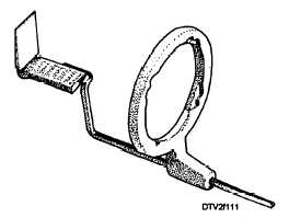
Figure 1-11. - Assembled anterior paralleling device.
Hold the base of the anterior bite-block between the thumb and first two fingers of your left hand. Ensure that the plastic film support is pointed upward and the film positioning slot is away from you.
Holding the film packet in position, press it against the plastic support and slide the film down into the positioning slot. The printed side of the packet should be facing the plastic support, and the raised dot should be located toward the positioning slot.
The two prongs of the indicator rod are inserted into the openings in the bite-block.
Slide the anterior locator ring onto the indicator rod. Look through the locator ring. If the bite-block and
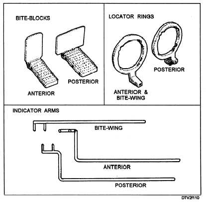
Figure 1-10. - Anterior and posterior paralleling devices.
Continue Reading