Seat and position the patient. Positioning varies according to the type of radiographic examination and the film placement technique you are going to use. Specific positioning procedures will be discussed later.
If the patient is a woman, ask her if she is pregnant. If she is or you suspect that she might be, consult the dentist.
Ask the patient to remove eyeglasses, complete dentures, removable partial dentures, earrings, or any other objects about the head and neck.
Explain the X-ray procedures to the patient. If the patient is nervous about being X-rayed, explain the safety precautions taken to prevent overexposure to radiation.
Drape the patient with a lead apron and thyroid collar.
Quickly examine the patient's mouth to determine its anatomy. Such things as a small mouth, an abnormally shallow vault, crooked teeth, and bony protrusions can affect the placement of the film packet. The patient's overall bone size and density will determine the kVp setting. For a patient with a normal bone size and density, use a kVp setting of 87; for a patient with a thick bone size and density, use a 90 kVp setting.
Position the patient's head securely against the headrest.
Place the film packet in the patient's mouth. Film placement procedures will be discussed later. Occasionally patients may gag when the film is placed in their mouth. The gagging reflex may be caused by nervousness, so remain calm and reassure the patient. You might recommend that patients breathe through their nose, since it is difficult to gag while doing so, having patients rinse out their mouth with cold water may also help or have patients concentrate on something other than gagging. Whatever technique you use you will have to be swift in placing the film and making the exposure because the chance of keeping the gag reflex from returning for an extended period is highly unlikely.
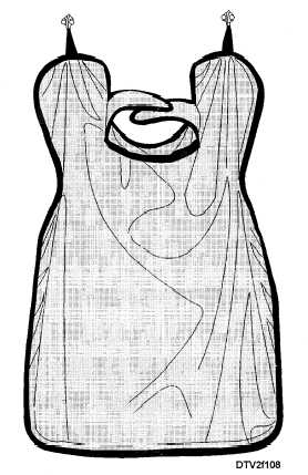
After the X-ray procedure is completed, you must store the lead apron and thyroid collar properly to avoid damage as shown in figure 1-8.
PERIAPICAL EXAMINATION
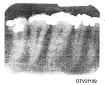
A periapical examination is conducted to obtain radiographs of the crowns, roots, and supporting

Figure 1-8. - Properly stored lead apron with thyroid collar.
structures of the teeth. Figure 1-9 shows a typical periapical radiograph.
There are two techniques available to take periapical radiographs: paralleling and bisecting- angle. Both techniques use the long axis of the tooth as a focal point. The paralleling technique is the preferred method and the bisecting-angle technique is used as an alternative. Film placement and techniques are discussed in the following sections.
PARALLELING TECHNIQUE
When using the paralleling technique, you must center the X-ray film packet behind, and parallel with

Figure 1-9. - Typical periapical radiograph.
Continue Reading