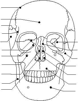
The maxillary bones form the upper jaw, the
anterior roof of the mouth, the floors of the orbits, and
the sides and floor of the nasal cavity. The small holes
on each side of the nasal opening are called the
infraorbital foramina (sing. foramen). The maxillary
bones contain large cavities called maxillary sinuses.
The palatine bones are L-shaped bones located
behind the maxillary bones. They form the posterior
section of the hard palate and the floor of the nasal
cavity.
The zygomatic bones are responsible for the
prominence of the cheeks. The zygomatic bones serve
as part of the posterior section of the hard palate and the
floor of the nasal cavity.
The lacrimal bones provide a pathway for a tube
that carries tears from the eye to the nasal cavity. The
lacrimal bone is a thin, scalelike structure located in
the medial wall of each orbit.
The nasal bones have cartilaginous tissues
a t t a c h e d t o t h e m . T h e s e t i s s u e s c o n t r i b u t e
significantly to the shape of the nose. The nasal bones
are long, thin, and nearly rectangular in shape. They lie
side by side and are fused together to form the bridge of
the nose.
The vomer bone is connected to the ethmoid bone,
and together they form the nasal septum (the wall
separating the two nasal cavities).
The middle and inferior nasal conchae are
fragile, scroll-shaped bones that are attached to the
lateral wall of the nasal cavity. The inferior nasal
concha provides support for mucous membranes
within the nasal cavity.
The lower jawbone is called the mandible. The
mandible is horseshoe-shaped with flat, bony
projections on each end. The two small holes on the
jawbone are called the mental foramina. The
mandible's main function is mastication (chewing
food).
VERTEBRAL (SPINAL) COLUMN.—The
vertebral column consists of 24 movable or true
vertebrae; the sacrum; and the coccyx, or tail bone (fig.
1-17). The vertebrae protect the spinal cord and the
nerves that branch out from the spinal cord. Each
vertebra has an anterior portion, called the body, which
is the large solid segment of the bone (fig. 1-18). This
vertebral body supports not only the spinal cord but
other structures of the body as well. At the bottom of
the spinal column is the sacrum and the coccyx. Many
of the main muscles are attached to the vertebrae.
The vertebral foramen is a hole directly behind
the body of the vertebrae that forms the passage for the
spinal cord. The vertebral projections are for the
attachments of muscles and ligaments and for
facilitating movement of one vertebra over another.
The spinal column is divided into five regions in the
following order: cervical (neck), thoracic (chest),
1-9
HM3F0116
PARIENTAL
BONE
FRONTAL
BONE
LACRIMAL
BONE
ETHMOID
BONE
SQUAMOSAL
SUTURE
TEMPORAL
BONE
SPHENOID
BONE
PERPENDICULAR PLATE
OF THE ETHMOID BONE
INFRAORBITAL
FORAMEN
VOMER
BONE
MANDIBLE
MENTAL
FORAMEN
MAXILLA
INFERIOR NASAL
CONCHA
ZYGOMATIC
BONE
MIDDLE NASAL
CONCHA
SPHENOID
BONE
NASAL
BONE
Figure 1-16.—Facial bones.