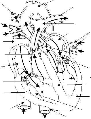
atrium and pumps it out into the arteries. The openings
between the chambers on each side of the heart are
separated by flaps of tissue that act as valves to prevent
backward flow of blood. The valve on the right has
three flaps, or cusps, and is called the tricuspid valve.
The valve on the left has two flaps and is called the
mitral, or bicuspid, valve. The outlets of the
ventricles are supplied with similar valves. In the right
ventricle, the pulmonary valve is at the origin of the
pulmonary artery. In the left ventricle, the aortic valve
is at the origin of the aorta. See figure 1-33 for valve
locations.
The heart muscle, the myocardium, is striated like
the skeletal muscles of the body, but involuntary in
action, like the smooth muscles. The walls of the atria
are thin with relatively little muscle fiber because the
blood flows from the atria to the ventricles under low
pressure. However, the walls of the ventricles, which
comprise the bulk of the heart, are thick and muscular.
The wall of the left ventricle is considerably thicker
than that of the right, because more force is required to
pump the blood into distant or outlying locations of the
circulatory system than into the lungs located only a
short distance from the heart.
Heart Functions
The heart acts as four interrelated pumps. The right
atrium receives deoxygenated blood from the body via
the superior and inferior vena cava. It pumps the
deoxygenated blood through the tricuspid valve to the
right ventricle. The right ventricle pumps the blood
past the pulmonary valve through the pulmonary
artery to the lungs, where it is oxygenated. The left
atrium receives the oxygenated blood from the lungs
through four pulmonary veins and pumps it to the left
ventricle past the mitral valve. The left ventricle
pumps the blood to all areas of the body via the aortic
valve and the aorta.
The heart's constant contracting and relaxing
forces blood into the arteries. Each contraction is
followed by limited relaxation or dilation. Cardiac
muscle never completely relaxes: It always maintains a
degree of tone. Contraction of the heart is called
systole or “the period of work.” Relaxation of the heart
is called diastole or “the period of rest.” A complete
cardiac cycle is the time from onset of one contraction,
or heart beat, to the onset of the next.
1-26
AORTIC ARCH
LEFT
PULMONARY
ARTERY
RIGHT
PULMONARY
ARTERY
PULMONARY
TRUNK
BRANCHES
OF LEFT
PULMONARY
VEIN
AORTIC
SEMILUNAR
VALVE
MITRAL
VALVE
LEFT
VENTRICLE
INTERVENTRICULAR
SEPTUM
MYOCARDIUM
(HEART MUSCLE)
PAPILARY
MUSCLE
DESCENDING
AORTA
INFERIOR VENA
CAVA
(FROM TRUNK
AND LEGS)
RIGHT
VENTRICLE
TRICUSPID
VALVE
RIGHT
ATRIUM
PULMONARY
SEMILUNAR
VALVE
SUPERIOR
VENA CAVA
(FROM HEAD
AND ARM)
FROM LUNG
FROM LUNG
TO LUNG
TO LUNG
LEFT
ATRIUM
HM3F0133
Figure 1-33.—Frontal view of the heart—arrows indicate blood flow.


