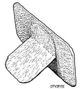
Figure 1-32. - Bitewing film packet.
PARALLEL PLACEMENT TECHNIQUE
The following procedures describe this technique:
1. Program the X-ray machine for the discussed time, mA settings, and kVp settings.
2. Prepare the inter-proximal paralleling device (fig. 1-33). Fold the bitewing tab against the film packet and insert the packet into the bite-block so that the printed side faces the backing support. Insert the end of the indicator rod into the holes in the bite-block. Slide the locator ring onto the indicator rod. Look through the locator ring to see if the bite-block is centered in the ring. If it is, the paralleling device is ready for positioning in the patient's mouth.
3. Position the paralleling device with film in the patient's mouth so that the anterior edge of the film touches the distal surface of the mandibular cuspid (fig. 1-34). Have the patient close gently but firmly on the bite-block to hold the film in position.
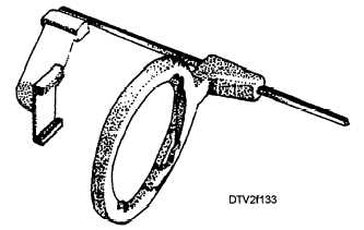
Figure 1-33. - Interproximal paralleling device.
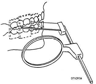
Figure 1-34. - Paralleling device positioned for interproximal radiographs.
4. Slide the locator ring down the indicator rod until the ring almost touches the surface of the patient's face. Then, align the tube head using the same technique as previously described for the paralleling device.
5. Make the exposure. After making the exposure, put the exposed film in a lead lined container or behind a protective screen. You are now ready to take the radiograph on the opposite side of the patient's mouth.
BISECTING-ANGLE TECHNIQUE
The following procedures describe this technique:
1. Program the X-ray machine for the discussed time, mA settings, and kVp settings.
2. Position the patient so that the ala-tragus line is parallel with the floor, and the midsagittal plane is perpendicular to the floor.
3. Position the film packet in the patient's mouth. Hold the wing of the packet between your thumb and index finger. Place the lower edge of the packet between the tongue and the lingual surfaces of the mandibular teeth. Position the packet so that its anterior edge touches the distal surface of the mandibular cuspid. Rest the wing of the packet on the occlusal surfaces of the mandibular teeth. Instruct the patient to close slowly. As the patient's maxillary teeth contact your index finger, roll your finger out facially, permitting the patient's teeth to close on the wing (fig. 1-35). The film packet is now positioned.
4. Set the vertical angulation of the tube head at +5° to +l0°.Continue Reading