2. Position the patient as shown in figure 1-23 for maxillary radiographs or figure 1-24 for mandibular radiographs. Remember that the patient's midsagittal plane must be perpendicular to the floor.
3. Position the film packet in the patient's mouth. Have the patient hold the film packet in place with a pair of hemostats or other holding device.
4. Set the vertical angulation of the tube head accord ing to the chart in figure 1-26.
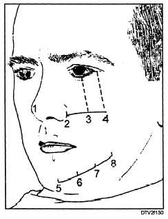
5. Center the tube head cylinder on the area to be radiographed. To simplify this process, the numbered anatomical landmarks are provided in figure 1-30. Take radiographs of the area by centering the tube head cylinder on these landmarks:
Maxillary incisor area: Landmark 1, the tip of the nose.
Maxillary cuspid area: Landmark 2, beside the ala of the nose.
Maxillary bicuspid area: Landmark 3, below the pupil of the eye.
Maxillary molar area: Landmark 4, below the outer angle of the eye and below the zygomatic bone.
Mandibular incisor area: Landmark 5, the tip of the chin.
Mandibular cuspid area: Landmark 6, directly below landmark 2 1/4 inches above the lower border of the mandible.

Figure 1-30. - Cylinder positioning landmarks for periapical radiographs.
Mandibular bicuspid area: Landmark 7, directly below landmark 3 1/4 inches above the lower border of the mandible.
Mandibular molar area: Landmark 8, directly below landmark 4 1/4 inches above the lower border of the mandible.
6. When you have the tube head cylinder centered on the horizontal landmark, double check to make sure that you have the correct horizontal angulation. The central X-ray beam should be projected straight through embrasures of the teeth to be radiographed.
7. Make the exposure.
8. Remove the film packet from the patient's mouth and place it in a clean paper cup. Place the disposable container in a lead container or behind a protective screen before making the next exposure.
INTERPROXIMAL (BITEWING) EXAMINATION
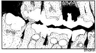
The interproximal examination reveals the presence of interproximal caries, certain pulp conditions, overhanging restorations, improperly fitting crowns, recurrent caries beneath restorations, and resorption of the alveolar bone.
A typical interproximal radiograph (fig. 1-31) records in a single exposure the coronal and cervical portions of both maxillary and mandibular teeth, along with the alveolar bone of the region.
Bitewing X-ray film packets are used for the interpromixal examination. The bitewing film packet (fig. 1-32) has a paper tab, or wing, that the patient bites on to hold the packet in place during the exposure (thus the name bitewing).
Interpromixal radiographs can be made using either the paralleling technique or the bisecting angle technique.

Figure 1-31. - Typical interproximal (bitewing) radiograph.
Continue Reading