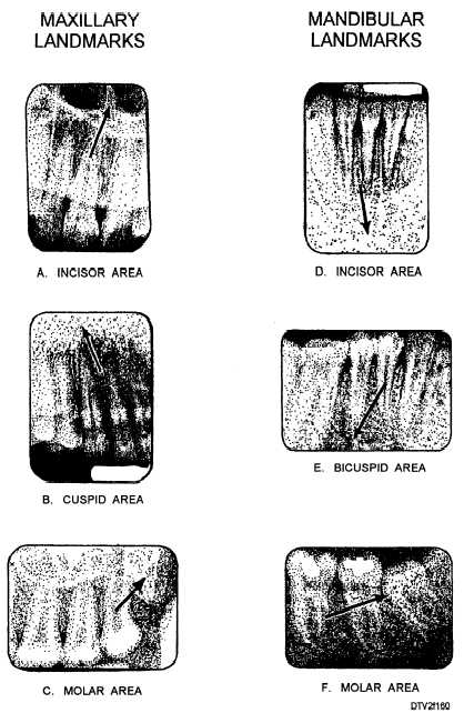
Figure 1-60. - Maxillary and mandibular radiographic landmarks.
MOUNTING PROCEDURES
Place all the radiographs in the full mouth periapical series on a dry, flat working surface with the dimple side up. On the front of the film mount, enter the patient's name, social security number, rank/rate, the date, and the name of the dental treatment facility. Place the mount face down on the working surface. The two small arrows on the back of the mount should point toward you. Follow these steps to mount the radiographs:
1. Check each radiograph and make sure that the surface with the raised dimple faces you.
2. Mount interproximal radiographs. If inter- proximal (bite-wing) radiographs are included in the full mouth series, insert them in the slots provided as previously discussed.
3. Divide the radiographs into maxillary and mandibular groups. Using the film viewer, locate the anatomical landmarks discussed earlier. The maxillary radiographs are inserted in the 7 slots across the top of the film mount and the mandibular radiographs in the 7 slots across the bottom.
4. Insert the maxillary radiographs. First, identify the radiograph of the central incisor area. Keeping the side with the raised dimple facing toward you, rotate theContinue Reading
