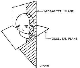
Figure 1-13. - Midsagittal and occlusal planes.
When taking a full mouth series or an individual periapical radiograph, follow the given guidelines for the specific area listed:
NOTE:
After each exposure, put the exposed film in a clean paper cup or disposable container. Then place the cup or disposable container in a lead container or behind a protective screen.
Maxillary Incisor Area
1. Set the exposure time selector to manufacturer's suggested impulses.
2. Prepare the anterior paralleling device.
3. Position the paralleling device with film in the patient's mouth. Center the film on the midline so that it is parallel with the long axis of the incisors (fig. 1-14).
4. Place a cotton roll under the bite-block. Have the patient close gently but firmly.
5. Adjust the locator ring and align the tubehead cylinder as previously described.
6. Make the exposure.
Maxillary Cuspid Area
1. Set the exposure time selector to manufacturer's suggested impulses.
2. Prepare the anterior paralleling device.
3. Position the paralleling device with film in the patient's mouth. Center the film on the cuspid and parallel with the tooth's long axis (fig. 1-15).
4. Place a cotton roll under the bite-block and have the patient close.
5. Adjust the locator ring and align the tubehead cylinder.
6. Make the exposure.
Maxillary Bicuspid Area
1. Set the exposure time selector to the manufacturer's suggested impulses.
2. Prepare the posterior paralleling device.
3. Position the paralleling device with film in the patient’s mouth. Center the second bicuspid and parallel with the tooth's long axis (fig. 1-16).
4. Place a cotton roll under the bite-block and have the patient close.
5. Adjust the locator ring and align the tubehead cylinder.
6. Make the exposure.
Maxillary Molar Area
1. Set the exposure time selector to the manufacturer's suggested impulses.
2. Prepare the posterior paralleling device.
3. Position the paralleling device with film in the patient's mouth. Center the film on the second molar, so the anterior edge of the film includes at least the distal half of the second bicuspid. The film should be parallel with the long axis of the molars (fig. 1-17).
4. lace a cotton roll under the bite-block and have the patient close.
5. Adjust the locator ring and align the tubehead cylinder.
6. Make the exposure.
Mandibular Incisor Area
1. Set the exposure time selector to the manu- facturer's suggested impulses.
2. Prepare the anterior paralleling device.
3. Position the paralleling device with film in the patient's mouth. Position the film packet so that it is centered on the midline and parallel with the long axis of the incisors (fig. 1-18).
4. Place a cotton roll on the upper surface of the bite-block and have the patient close.
5. Adjust the locator ring and align the tubehead cylinder.
6. Make the exposure.Continue Reading
