A large carious lesion with a pulpal exposure
Blood or pus oozing from the pulpal exposure
A fractured tooth or missing restoration
Treatment
As part of the emergency treatment plan for acute pulpitis, you may need to perform some of the following procedures:
Perform emergency treatment guidelines.
Gently remove loose debris from the cavity.
Dry the cavity with cotton pellets.
Pack the cavity with a cotton pellet slightly moistened with eugenol.
Gently fill the cavity with a temporary filling material.
Check the occlusion.
Instruct the patient to return for definitive treatment.
PERIAPICAL ABSCESS
A periapical abscess (fig. 6-4) usually results from an infection of the pulpal tissue causing the pulp to become necrotic (die). This type of infection causes fluids and by-products to build up within the walls of the pulp chamber and root canal(s). The periapical abscess is formed when these materials escape through the apical foramen of the tooth. An area of pus and fluid accumulation forms in the bone surrounding the apex of the tooth. As the pressure builds up, a channel may form through the alveolar bone and the soft tissue. This channel is called a sinus tract. When the pus reaches the soft tissue, vestibular or facial swelling can occur. Extensive swelling is called cellulitis. Swelling that is confined to a small area at the site of a sinus tract is called a gumboil.
Symptoms
A patient with periapical abscess may complain of the following:
Constant, throbbing pain in the affected area.
Increased pain when chewing.
Increased pain when lying down.
Bad taste in the mouth.
A gumboil. The tooth "feels longer" than the others.
Malaise.
Tender lymph nodes.
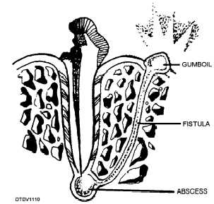
Figure 6-4. - Periapical abscess.
Signs
When there is a periapical abscess, you may observe some of the following signs upon examination:
A severe pain reaction is experienced when light pressure is applied to the affected tooth.
A gumboil.
Facial swelling (general or localized).
Tooth mobility.
An elevated temperature.
Enlarged lymph nodes.
Treatment
To treat the periapical abscess, you should perform the following in the emergency treatment plan:
Perform emergency treatment guidelines.
Expose a periapical radiograph of the affected tooth. The abscess will appear as a radiolucency around the apex of the tooth.
Drain the abscess. If the abcess is soft and pus is evident, drainage can be done without local anesthesia. Puncture the most raised portion of the abscess with an explorer.
If a carious lesion is present, gently excavate the cavity. NOTE: If drainage occurs through the cavity, the patient may experience a rapid relief from pain.Continue Reading