affected tooth can manifest in a healthy, noninvolved tooth; this is called referred pain.
Signs
Upon examination you may find the following signs of an infection:
A chalky white spot on the enamel
Roughness on the surface of the tooth
A dark, stained cavity
A cavity filled with food or a spongy mass of decaying dentin
Treatment
As a part of the emergency treatment plan, you may perform the following duties:
Perform emergency treatment guidelines.
Gently remove all debris from the cavity with a spoon excavator as illustrated in figure 6-1.
Flush the cavity with warm water.
Isolate the tooth with cotton rolls or gauze.
Carefully dry the cavity with cotton pellets as illustrated in figure 6-2.
Mix a temporary filling (zinc oxide eugenol, IRM, etc.).
Gently fill the cavity with the temporary filling material as illustrated in figure 6-3.
Check the occlusion. Make sure the temporary restoration does not touch the opposing tooth.
Instruct the patient to return for definitive treatment on the next work day.
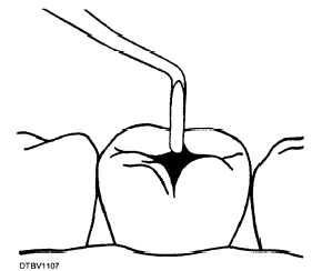
Figure 6-1.—Removing debris from the cavity.
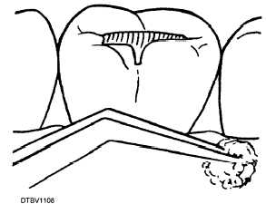
Figure 6-2.—Preparing to dry the cavity.
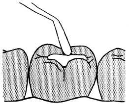
Figure 6-3.—Placing the temporary filling.
ACUTE PULPITIS
Acute pulpitis is an inflammation of the pulp caused by injury to the pulp, usually from dental caries or trauma. It is the most frequent cause of severe tooth pain. The pain is caused by the pressure of fluids building up inside the pulp chamber or root canal.
Symptoms
A patient with acute pulpitis may complain of the following:
Spontaneous, continuous, or intermittent pain that lingers
Piercing and pulsating pain in the affected area
Increased pain when lying down
Signs
Upon examination for acute pulpitis, you may observe one of the following signs:
A large carious lesionContinue Reading