floor of the orbits are the maxillary sinuses; the largest of the sinuses.
Palatine Bones
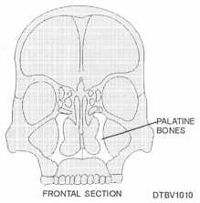
The palatine bones are located behind the maxillae (fig. 3-10). The bones are somewhat L-shaped and form the posterior portion of the hard palate and the floor of the nose. Anteriorly, they join with the maxillary bone.
Zygomatic Bones (Zygoma, Malar Bone)
The zygomatic bones make up the prominence of the cheeks and extend from the zygomatic process of the temporal bone to the zygomatic process of the maxilla. The zygomatic bones form the "cheek bones" and help to form the sides and floor of the orbits.

Figure 3-10. - Anterior view of palatine bones.
Lacrimal Bones
The lacrimal bones are the smallest and most fragile of the cranial bones. These thin, scalelike structures are located in back of the frontal process of the maxilla.
Nasal Bones
The nasal bones are small oblong bones somewhat rectangular in shape. They lie side by side and are fused at the midline to form the bridge of the nose (nasal septum). These bones are responsible for the shape of the nose.
Inferior Nasal Conchae
The inferior nasal conchae are curved, fragile, scroll-shaped bones that lie in the lateral walls of the nasal cavity. They provide support for mucous membranes within the nasal cavity.
Vomer Bone
The vomer bone is a thin, flat, single bone almost trapezoid in shape. It connects with the ethmoid bone and together they form the nasal septum.
Mandible
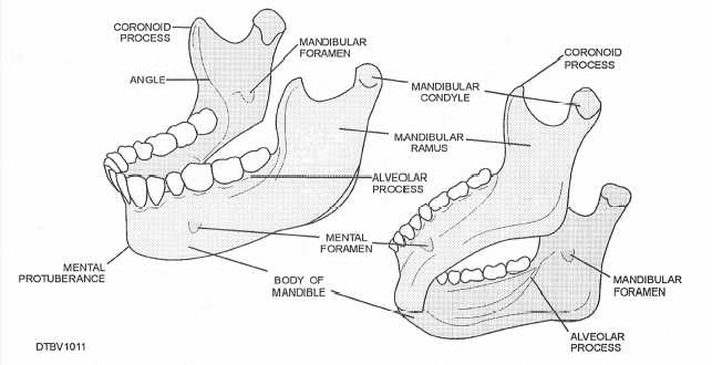
The mandible (lower jaw-bone) is the longest, strongest, and the only movable bone in the skull. Figure 3-11 illustrates the anatomy of the mandible.

Figure 3-11. - Anatomy of the mandible; lateral view (left), inferior view (right).
Continue Reading