The mandible is horseshoe-shaped, with an upward sloping portion at each end called the ramus. The rami are divided into two different processes:
Condyloid process-Also called mandibular condyle, located posterior on the ramus and forms the head of the mandible. It is knuckle-shaped, and articulates in the glenoid fossa of the temporal bone to form the temporal mandibular joint.
Coronoid process-Located anterior of the condyle, and provides attachment for the temporal's muscle, which helps lift the mandible to close the mouth.
Other important anatomical landmarks of the mandible you should be able to recognize are as follows:
Alveolar process-Supports the teeth of the mandibular arch.
Mental protuberance-Also referred to as the chin and is located at the midline of the mandible.
Mental foramen-Located on the facial surfaces of the mandible on both the right and left sides, just below the second premolars. This opening contains the mental nerve and blood vessels.
Body-The heavy, horizontal portion of the mandible below the mental foramen extending from the angle to the parasyplysis region.
Angle-Juncture where the body of the mandible meets with the ramus.
Mandibular foramen—Located near the center of each ramus on the medial side (inside), through this opening passes blood vessels and the interior alveolus nerve, which supply the roots of the mandibular teeth. This is a common area where the dental officer will inject anesthetic to block the nerve impulses and make the teeth on that side insensitive (numb).
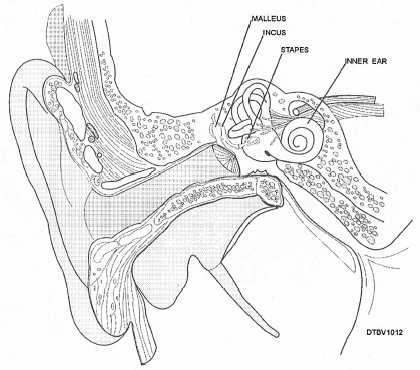
BONES OF THE EAR
In each middle' ear and located in the auditory ossicles are three small bones named the malleus, incus, and staples (fig. 3-12). Their function is to transmit and amplify vibrations to the ear drum and inner ear.

Figure 3-12. - Anatomy of the middle ear.
TEMPORAL MANDIBULAR JOINT
The right and left temporal mandibular joints (TMJs) are formed by the articulation of the temporal bone and the mandible. This is where TMJs connect with the rest of the skull. Figure 3-13 illustrates the TMJ.
The mandible is joined to the cranium by ligaments of the temporal mandibularjoint (fig. 3-14).
Continue Reading