areas of tenderness or swelling in the vaginal walls. Identify the cervix and note its position, consistency, mobility, and indications of cervical tenderness on motion. Palpate the fornix as illustrated in figure 2-4 (I). Using your other hand (referred to as the abdominal hand), palpate downward midway between the umbilicus and the symphysis pubis toward your pelvic hand. Identify the uterus between your hands, noting 159.35
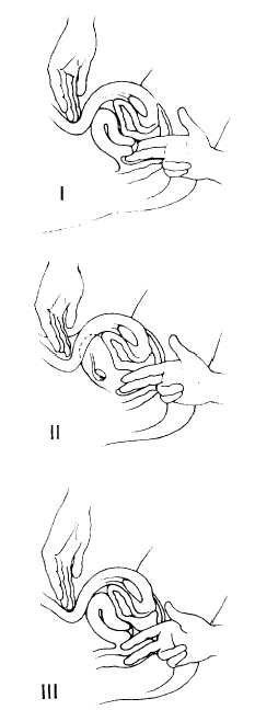
Figure 2-4.-Techniques of bimanual examination.
any masses or tenderness, the size, shape, consistency, and mobility (fig. 2-4 (II)). Place your pelvic hand in the right lateral fornix and your abdominal hand in the right lower abdominal quadrant. Exert downward pressure with your abdominal hand and palpate the ovary. Note the size, shape, consistency, and presence of any masses or tenderness. Repeat the procedures for the left side.
Withdraw your fingers from the vagina. Relubricate, if necessary, and then slowly introduce your middle finger into the patient’s rectum and your index finger into her vagina (fig. 2-4 (III)). The anal sphincter may be relaxed by asking the patient to bear down while you are introducing your fingers. Repeat the steps of the bimanual examination. Pay special attention to the region that lies behind the cervix and the posterior uterine surface itself, as these areas may only be accessible to the rectal finger. Take note of any masses or areas of tenderness. Look for signs of rashes, excoriation, and external hemorrhoids.
COMMONLY ENCOUNTERED FEMALE CONDITIONS
Vaginitis This is an inflammation of the vaginal mucosa caused by fungal, bacterial, or mechanical factors. The zone of inflammation may extend from the vagina to the cervix and the vulvar region. It may be caused by inflammations of Bartholin’s or Skene’s gland ducts. The three most commonly encountered forms of vaginitis are Trichomonas, Monilia, and bacterial.
SYMPTOMS—The most prominent symptom will be leukorrhea. With this type of vaginitis, the discharge may be thick or thin and profuse, may have a fetid odor, and will range in color from white to greenish-yellow. The discharge is often pooled in the vaginal fornix and is quite often bubbly in appearance. Visualization of the vaginal mucosa will disclose a red, inflamed mucosa and a cervix with small red, granular, strawberry looking spots. The patient will normally reveal a history of vulvar (external genitalia) irritation, vaginismus (painful spasms of the vagina), dyspareunia (painful coitus), and itching. Motile