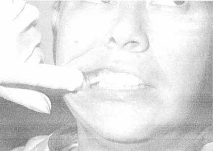
Figure 1-35. - Patient closing on wing.
5. Center the tube head cylinder on the wing of the film packet. Be sure that the central X-ray beam passes through the embrasures as shown in figure 1-36.
6. Make the exposure. After making the exposure, put the exposed film in a clean paper cup and place in a lead lined container or behind a protective screen. You are now ready to take the radiograph on the opposite side of the patient's mouth.
OCCLUSAL EXAMINATION
An occlusal examination is usually conducted when fractures of the jaw or gross pathological conditions are suspected. A typical occlusal radiograph (fig. 1-37) shows a large area of the maxillary or mandibular arch.
The occlusal film packet is shaped much like the periapical packet, only larger. Unlike the periapical
Figure 1-36. - Centering tube head cylinder.
Continue Reading