RADIATION SAFETY
Proper safety precautions must be observed by all persons working in or near an area where X-rays are being generated. X-rays can be dangerous. Long term overexposure to radiation may result in loss of hair, redness and inflammation of the skin, blood count change, cell atrophy (wasting away), ulcerations, sterility, genetic damage, cancer, leukemia, and death.
There are safety measures designed to protect the patient and the health care team from the dangers of overexposure to radiation and the operation of X-ray equipment. You must observe these safety measures when working in radiology. Your command will have instructions and standard operating procedures (SOP) for the operation of dental radiographic (X-ray) units and equipment. You will be required to read these procedures if you are newly assigned to the radiology department. There are other numerous responsibilities that include providing radiology support for oral diagnosis, log maintenance, infection control, testing for quality control, and processor maintenance.
PATIENT PROTECTION
A number of precautions are taken to prevent the patient from being exposed to inappropriate diagnostic radiation. The decision to order dental radiographs is determined by the dental officer on a case by case basis for each patient. Only a dental officer is authorized to order and diagnostically interpret dental radiographs.
Perhaps the most important safety measure is the responsibility of the assistant: When taking radiographs, you should always have patients wear lead aprons and thyroid collars to shield their reproductive organs and thyroid glands. There is only one exception to this rule; when obtaining a panorex radiograph, the thyroid collar is not used since it blocks part of the X-ray beam. In addition, always ask a female patient whether or not she is pregnant or if pregnancy is questionable, before taking radiographs. If she is pregnant, consult the dental officer.
Other radiation safety measures include X-ray machines that have built-in safeguards that filter out harmful radiation and restrict the central X-ray to the smallest possible area. Fast film is used to shorten expos ure time; and only essential radiographs are taken on patients.

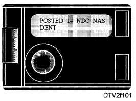
Figure 1-1. - Environmental dosimetry radiation film badge.
ASSISTANT PROTECTION
When you work near a source of radiation, your X-ray department will be issued an environmental dosimetry radiation film badge (fig. 1-1). feet from the tube head and never in the direct line of radiation during expos ure. These film badges
Appropriately placed environmental film badges are used to monitor stray radiation that may occur in and around the X-ray department. The badges are placed in the X-ray room behind the technicians protective lead-lined barrier or at least 6 contain X-ray sensitive film in a light-tight packet. The film packets are collected every 6 to 7 weeks. After collection, the film is sent to the radiation detection laboratory for processing and evaluation. Any abnormally high readings (i.e., greater than 0.010 REM [Radiological Equivalent Mammel]) shall be referred to the Radiation Health Office for investigation.
When you take radiographs on a patient, observe the following precautions to avoid unnecessary exposure to radiation:
NEVER stand in the path of the central X-ray beam during exposure.
NEVER hold the X-ray film packet in the patient's mouth during exposure.
NEVER hold the tube head or the tube head cylinder of the X-ray machine during exposure.
ALWAYS stand behind a lead-lined screen during an exposure.
X-RAY FILM LOG
Another portion of radiation safety is to account for all radiographs that are taken. An X-ray film log
Continue Reading