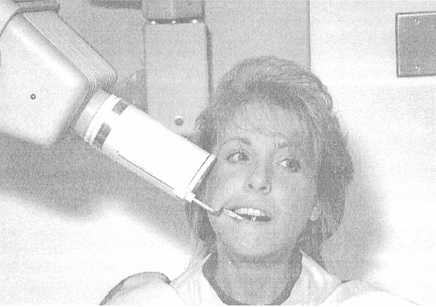

Figure 1-17. - Film and cylinder placement: maxillary molar area.
distance of 8 inches (short cone).
The bisecting-angle technique is not recommended for routine use.
Since paralleling devices are not used with the bisecting-angle technique, you must pay special attention to positioning the patient, the film packet, and the tube head.
Positioning the Patient
For all maxillary periapical radiographs, position the patient's head as shown in figure 1-23 from the ala of the nose (the outer portion of the nostril) to the tragus of the ear (a projection of the cartilage on the front center of the ear). This ala-tragus line should be parallel with the floor. The patient's head should also be positioned so that the midsagittal plane is perpendicular to the floor.
For mandibular periapical radiographs, lower the headrest so the patient's head is positioned as shown in figure 1-24. The figure shows a line running from the corner of the patient's mouth to the tragus of the ear. This line should be parallel with the floor. Again, the mid-sagittal plane is perpendicular to the floor.
Positioning the Film
Once the patient is positioned, insert the film packet in the patient's mouth with a pair of hemostats or other holding device. Never slide the packet in; this might irritate the oral mucosa or cause the patient to gag. Gently direct the holding device to the desired
Continue Reading