Marginal Gingivitis
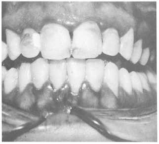
Marginal gingivitis (fig. 5-7) is the most common type of gingival disease. Most frequently it is the result of poor oral hygiene and affects both the gingival margins and papilla. Chief irritants are food debris and plaque around the necks of the teeth, interproximal spaces, or overhanging margins of dental restorations. Occasionally, a localized inflamed condition may exist from a popcorn husk or toothbrush bristle. Early formation of calculus deposits can also form under the gingival sulcus (subgingival) on the facial and lingual surfaces of the upper and lower teeth. Calculus deposits can also be responsible for the occurrence of marginal gingivitis, and if left untreated, may proceed to destruction of the supporting structures (as in periodontitis).
Marginal gingivitis usually starts at the tips of the papillae and then extends to the gingival margins.
Swelling, loss of stippling (orange peel texture of surface) of the attached gingiva, redness, easily retractable sulcus, and foremost, a tendency to bleed easily, are the main characteristics. This condition may be generalized (exist around all teeth), or it may be localized to one or two or a group of teeth.
Acute Necrotizing Ulcerative Gingivitis
Acute necrotizing ulcerative gingivitis (ANUG) (fig. 5-8) is a disease commonly referred to as trenchmouth, or Vincent’s infection. It is characterized during the acute stage by redness, swelling, pain, accumulation of calculus around the sulcus of the teeth, and bleeding of the gingival tissues. Usually there is a film of necrotic white or grayish tissue around the teeth. This membrane may be wiped off, leaving a raw, bleeding base. The ulceration of the gingival crest results in a characteristic punched-out appearance and loss of the interdental papillae. There is an unpleasant odor and a foul taste in the mouth. The gingival tissues bleed easily when touched, and patients will complain of not being able to brush their teeth or chew well because of the pain or discomfort.
PERIODONTITIS
Periodontitis (fig. 5-9) is a chronic inflammatory condition that involves the gingiva, crest of the alveolar bone, and periodontal membrane. This condition results in loss of bone that supports the teeth, periodontal pocket formation, and tooth mobility. It usually develops as a result of untreated chronic marginal gingivitis. The color of the gingival tissues is intensified and becomes bluish red as the disease progresses. A gradual recession of the periodontal tissue will occur. Neglected deposits of calculus and formation of additional calculus over time contribute to the spread of the disease. Like marginal gingivitis, it may affect the entire dentition, or only localized areas.

Figure 5-7. - Marginal gingivitis.
Continue Reading