Table 3-4. - Muscles of Mastication
Muscle
Origin Masseter Zygomatic arch Temporalis Temporal bone
Insertion Mandible (external surface) Coronoid process at the anterior border of the ramus
Description Closes jaw; flat, thick muscle Closes jaw; fan-shaped Medial pterygoid Lateral pterygoid Sphenoid, palatine, and maxillary bones Sphenoid bone Inner (medial) surface of Closes jaw; parallels the ramus masseter muscle Anterior surface of man- Opens jaw; allows dibular condyle grinding action side to side, and protrudes the mandible reception. We receive food in the mouth, reducing it in size, and mixing it with saliva for the digestion process.
CHEEKS
The cheeks are the side walls of the mouth. They are made up of layers of skin, a moist inner lining called mucosa, fat tissue, and certain muscles. The buccinator muscle of the cheeks prevents food from escaping the chewing action of the teeth.
LIPS The lips are covered externally by skin and internally by the same mucous membranes that line the oral cavity. They form the anterior border of the mouth. The area of the external lips where the red mucous membrane ends and normal outside skin of the face begins is known as the vermilion border. Figure 3-17 illustrates the anatomy of the lips.
The lips are very sensitive and act as sensory receptors, allowing food and liquids to be placed in the
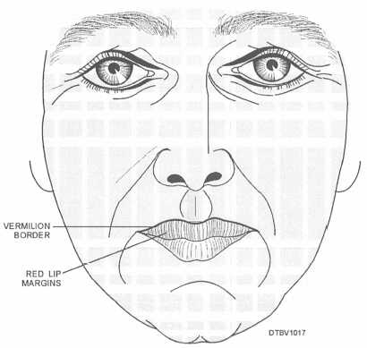
Figure 3-17. - Anatomy of the lips.
Continue Reading