ocular muscles, eyelids, conjunctival, and lacrimal apparatus.
Table 3-1.—Functions of the autonomic nervous system
| Sympathetic | Parasympathetic |
| Dilates pupils. | Constricts pupils. |
| Lessens tonus of ciliary muscles so the eyes may accommodate to see distant objects. | Contracts ciliary muscles so the eyes may accommodate to see objects near at hand. |
| Dilates bronchi. | Constricts bronchi. |
| Quickens and strengthens the action of the heart. | Slows the action of the heart. |
| Contracts blood vessels of the skin and viscera so that more blood goes to the skeletal and cardiac muscles where it is needed for “fight or flight.” | Dilates blood vessels (except cardiac). |
| Relaxes gastrointestinal tract and bladder. | Increases contraction of gastrointestinal tract and muscle tone of the bladder. |
| Decreases secretions of the gastrointestinal glands. | Increases secretions of gastrointestinal glands. |
| Increases secretion of sweat glands. | No action on sweat glands. |
| Causes contraction of sphincters to prevent emptying of bowels or bladder. | Relaxes sphincters so that waste matter can be excreted. |
Structure of The Eye
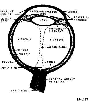
The eye is a hollow ball, or globe, which consists of various tissues that perform specific functions. The globe, or eyeball, is composed of three layers (fig. 3-45).

Figure 3-45.—Cross section of the eye.
OUTER LAYER.—The outer layer of the eye is called the sclera. It is the tough, fibrous, protective portion of the globe, commonly called the white of the eye. Anteriorly, the outer layer is transparent and is called the cornea, or the window of the eye. It permits light to enter the globe. The exposed sclera is covered with a mucous membrane, the conjunctival, which is a continuation of the inner lining of the eyelids. The lacrimal gland produces tears that constantly wash the front part of the eye and the conjunctival. The tear gland secretions that do not evaporate flow
