myelin sheath), causing them to appear gray. Beneath this layer is the medulla. This is often called the white matter of the brain, because the nerves are myelinated (covered with a myelin sheath and an outer covering called the neurilemma), which gives them their white appearance (fig. 3-40).
The cortex of the cerebrum is irregular. It bends on itself in folds called convolutions, which are separated from each other by grooves and fissures. The deep sagittal cleft, a longitudinal fissure, divides the cerebrum into two hemispheres. Other fissures further subdivide the cerebrum into lobes, each of which serves a localized, specific brain function (fig. 3-41). For example, the frontal lobe is associated with the higher mental processes such as memory, the parietal lobe is concerned primarily with general sensations, the occipital lobe is related to the sense of sight, and the temporal lobe is concerned with hearing.
The cerebellum is situated posteriorly to the brain stem, which is made up of the pens, midbrain, and medulla oblongata, and inferior to the occipital lobe. It is concerned chiefly with bringing balance, harmony, and coordination to the motions initiated by the cerebrum.
Two smaller divisions of the brain, vital to life, are the pens and the medulla oblongata. The pens consists chiefly of a mass of white fibers connecting the other three parts of the brain—the cerebrum, cerebellum, and medulla oblongata.
The medulla oblongata is the inferior portion of the brain, the last division before the beginning of the spinal cord. It connects to the spinal cord at the upper level of the first cervical vertebra (C-1). In it are the centers for the control of heart action, breathing, circulation, and other vital processes such as blood pressure.
The outer surface of the brain and spinal cord is covered with three layers of membranes called the meninges. The dura mater is the strong outer layer; the arachnoid membrane is the delicate middle layer; and the pia mater is the vascular innermost layer that adheres to the surface of the brain and spinal cord. Inflammation of the meninges is called meningitis. The type depends upon whether the brain, spinal cord, or both are affected.
Cerebrospinal fluid is formed by a plexus (network) of blood vessels in the central ventricles of the brain. It is a clear, watery solution similar to blood plasma. The total quantity bathing the spinal cord is about 75 ml. It is constantly being produced and reabsorbed. It circulates over the surface of the brain and spinal cord and serves as a protective cushion as well as a means of exchange for nutrients and waste materials.

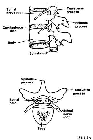
Figure 3-42.—Spinal cord.
Spinal Cord
The spinal cord is continuous with the medulla oblongata and extends from the foramen magnum, down inside the atlas, to the lower border of the first lumbar vertebra, where it tapers to a point. The cord is surrounded by the bony walls of the vertebral canal (fig. 3-42). It is unsheathed in the three protective meninges and surrounded by adipose tissue and blood vessels. The cord does not completely fill the vertebral canal, nor does it extend the full length of it. The nerve roots serving the lumbar and sacral regions must pass some distance down the canal before making their exit.
