The hip, or INNOMINATE, is a large irregularly shaped bone composed of three parts: the ilium, ischium, and pubis. In children these three parts are separate bones, but in adults they are firmly united to form a cuplike structure, called the ACETABULUM, into which the head of the femur fits. The ILIUM forms the outer prominence of the hip bone (crest of the ilium), the ISCHIUM forms the hard lower part, and the PUBIS forms the front part of the pelvis.
The area where the two pubic bones meet is called the SYMPHYSIS PUBIS and is often used in anatomical measurements. The largest foramen (opening) is located in the hip bone, between the ischium and the pubis, and is called the OBTURATOR FORAMEN. The crest of the ilium is used in making anatomical and surgical measurements (e.g., location of the appendix, which is approximately halfway between the crest of the ilium and the umbilicus).
The FEMUR, or thigh bone, is the longest bone in the body. The proximal end is rounded and has a head supported by a constricted neck that fits into the acetabulum. Two processes called the GREATER and LESSER TROCHANTERS are at the proximal end for the attachment of muscles. The neck of the femur, located between the head and the trochanters, is the site most frequently fractured. At the distal end are two bony prominences called the LATERAL and MEDIAL CONDYLES, which articulate with the tibia and the patella.
The PATELLA is a small oval-shaped bone overlying the knee joint. It is enclosed within the tendon of the quadriceps muscle of the thigh. Bones like the patella that develop within a tendon are known as SESAMOID bones.
The TIBIA, or shin bone, is the larger of the two leg bones and lies at the medial side. The proximal end articulates with the femur and the fibula. Its distal end articulates with the talus (one of the foot bones) and the fibula. A prominence easily felt on the inner aspect of the ankle is called the MEDIAL MAL.LEOLUS.
The FIBULA, the smaller of the two leg bones, is located on the lateral side of the leg, parallel to the tibia. The prominence at the distal end forms the outer ankle, known as the LATERAL MALLEOLUS.
The TARSUS, or ankle, is formed by seven tarsal bones. The strongest of these is the heel bone or CALCANEUS.
The sole and instep of the foot is called the METATARSUS and is made up of five METATARSAL bones. They are similar in arrangement to the metacarpal of the hand.
The PHALANGES are the bones of the toes and are similar in number, structure, and arrangement to the bones of the fingers.
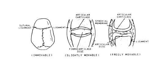
Figure 3-23.—Typical joints.
JOINTS
Whenever two bones are attached to each other, a joint is formed. In a freely movable joint, such as the knee or elbow joint, the ends of the bones are covered with a smooth layer of cartilage. The whole joint is enclosed in a watertight sac or membrane containing a small amount of lubricating fluid. This enables the joint to work with little friction. The ligaments that reach across the joints from one bone to another keep them from getting out of place. When ligaments are accidentally torn, we call the injury a sprain; when bones are out of place, there is a dislocation; and when bones are chipped or broken, the injury is called a fracture.
Joints are classified according to the amount of movement they permit (fig. 3-23). They may be:
1. IMMOVABLE. Bones of the skull are rigidly interlocked along immovable joint lines known as sutures.