FEMALE REPRODUCTIVE SYSTEM
The female reproductive system (fig. 3-56) includes the ovaries, the fallopian (uterine) tubes, the uterus, the vagina, the external genitalia (vulva), and the breasts (mammary glands), which are not illustrated but will be discussed.
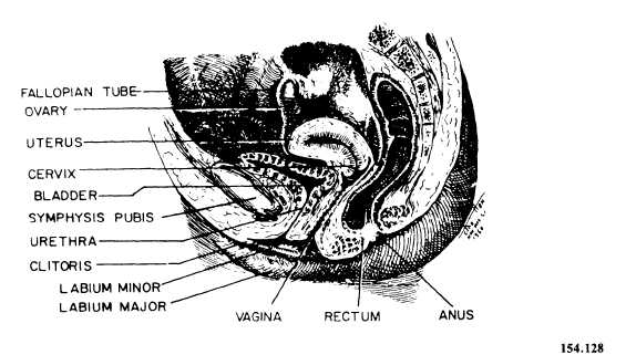
Figure 3-56.—The female reproductive system.
EXTERNAL GENITALIA
The external genital organs, referred to collectively as the vulva, include the mons pubis, labia majora, labia minors, clitoris, vestibule, Bartholin’s glands, and hymen. The mons pubis is the pad of fatty tissue beneath the skin, anterior to the symphysis pubis. The labia majora are two folds of skin extending from the mons pubis anteriorly to the perineum (the region between the vaginal orifice and the anus). Within these two folds of skin are two smaller folds, called the labia minors, extending from the clitoris to either side of the vaginal orifice. The clitoris is a small body richly endowed with nerves, highly sensitive, and of significance in sexual stimulation. The clitoris becomes engorged with blood during sexual excitement, but, unlike its male counterpart, the penis, it does not become erect. It is located at the point where the two labia minors meet. The vestibule is the area between the labia minors into which the urethral and vaginal orifices open. The urinary meatus is the external urethral orifice situated inferior to the clitoris and superior to the vaginal orifice. The vaginal orifice is situated inferior to the urethra. The Bartholin’s glands are the female counterparts of the Cowper’s glands in the male. They consist of two small roundish bodies on either side of the vaginal opening. Each gland is connected with the vagina by means of long ducts and secretes a viscid, alkaline fluid lubricant between the labia minors and the hymen. Finally, the hymen is a fold of mucous membrane that extends across the lower part of the vagina. It is not a very reliable indicator of virginity.
MAMMARY GLANDS
The mammary glands, or breasts, are accessory organs of the female reproductive system. They develop during puberty under the influence of the hormones estrogen and progesterone. The breasts are responsible for the secretion of milk (lactation) for the nourishment of newborn infants. Structurally the breasts resemble sweat glands. At the center is a nipple containing 15 to 20 depressions, into which ducts from the lobes of the gland empty. During pregnancy hormones secreted by the ovaries cause the glandular tissue to grow in preparation for lactation. After childbirth hormones secreted by the anterior lobe of the pituitary gland stimulate production for 6 to 9 months.