(fig. 1-41). The lungs are cone-shaped organs that lie
in the thoracic cavity. Each lung contains thousands of
alveoli with their capillaries. The right lung is larger
than the left lung and is divided into superior, middle,
and inferior lobes. The left lung has two lobes, the
superior and the inferior.
Pleurae
The pleurae are airtight membranes that cover the
outer surface of the lungs and line the chest wall. They
secrete a serous fluid that prevents friction during
movements of respiration.
Mediastinum
The mediastinum is the tissue and organs of the
thoracic cavity that form a septum between the lungs. It
extends from the sternum to the thoracic vertebrae and
from the fascia of the neck to the diaphragm. The
mediastinum contains the heart, the great blood
vessels, the esophagus, a portion of the trachea, and the
primary bronchi.
Diaphragm
The diaphragm is the primary muscle of
respiration. It is a dome-shaped muscle and separates
the thoracic and abdominal cavities. Contraction of
this muscle flattens the dome and expands the vertical
diameter of the chest cavity.
Intercostal Muscles
The intercostal muscles are situated between the
ribs. Their contraction pulls the ribs upward and
outward, resulting in an increase in the transverse
diameter of the chest (chest expansion).
Inhalation is the direct result of the expansion
caused by the action of the diaphragm and intercostal
muscles. The increase in chest volume creates a
negative (lower than atmospheric) pressure in the
pleural cavity and lungs. Air rushes into the lungs
through the mouth and nose to equalize the pressure.
Exhalation results when the muscles of respiration
relax. Pressure is exerted inwardly as muscles and
1-35
HM3F0140
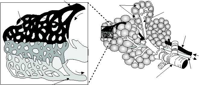
ALVEOLI
ALVEOLAR
DUCT
PULMONARY
ARTERY
BLOOD
FLOW
BRONCHIOLE
PULMONARY
VEIN
PULMONARY ARTERIOLE
CAPILLARY NETWORK
ON SURFACE OF ALVEOLUS
BLOOD
FLOW
BLOOD
FLOW
PULMONARY VENULE
Figure 1-40.—Bronchiole and alveoli.
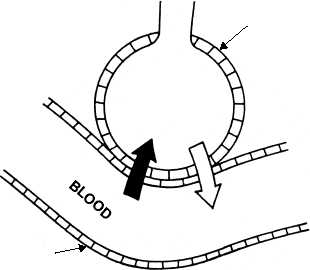
HM3F0141
ALVEOLAR
WALL
ALVEOLUS
PULMONARY
CAPILLARY
O2
CO2
Figure 1-41.—Pulmonary exchange at alveolus.