Lumbar.—There are five lumbar vertebrae.
Located in the small of the back, these vertebrae are the
larger and stronger segments of the vertebral column.
Sacrum.—The sacrum is the triangular bone
immediately below the lumbar vertebrae. It is
composed of five separate vertebrae that gradually
fuse together between 18 and 30 years of age. The
sacrum is connected on each side with the hip bone and
with the coccyx to form the posterior wall of the pelvis.
THORAX.—This cone-shaped bony cage is
about as wide as it is deep (fig. 1-20). The thorax is
formed by 12 ribs on each side and articulates
posteriorly with the thoracic vertebrae. The first set of
ribs are attached to the manubrium, a flat irregular
bone atop the sternum. The first seven pairs of ribs are
called true ribs. The remaining five pairs are called
false ribs. They are called false ribs because their
cartilages do not reach the sternum directly. The
eighth, ninth, and tenth ribs are united by their
cartilages and joined to the rib above. The last two rib
pairs, also known as floating ribs , have no
cartilaginous attachments to the sternum. The
sternum is an elongated flat bone, forming the middle
portion of the upper half of the chest wall in front. The
xiphoid process, located at the inferior aspect of the
sternum, serves as a landmark in the administration of
cardiopulmonary resuscitation.
1-11
HM3F0118
SPINOUS
PROCESS
LAMINA
TRANSVERSE
PROCESS
SUPERIOR
ARTICULAR
PROCESS
PEDICLE
BODY
PEDICLE
BODY
INTERVERTEBRAL
FORAMEN
INFERIOR
ARTICULATING
PROCESS
SUPERIOR
ARTICULATING
PROCESS
TRANSVERSE
PROCESS
FACET FOR
TUBERCLE
OF RIB
SPINOUS
PROCESS
A
B
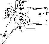
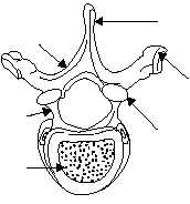
Figure 1-18.—Typical vertebra: A. Lateral view of a typical
vertebra; B. Superior view of a typical thoracic vertebra.
INTERVERTEBRAL
FORAMEN
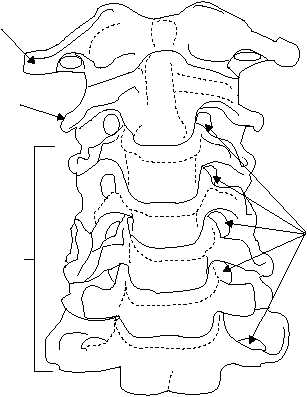
3RD TO 7TH
CERVICAL
VERTEBRAE
HM3F0119
AXIS
ATLAS
Figure 1-19.—Cervical vertebrae.