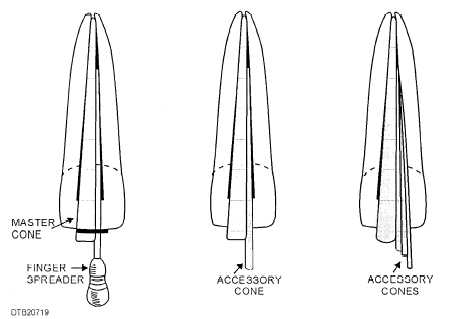
Figure 7-19. - Steps in filling a root canal with master and accessory cones.

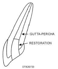
Figure 7-20. - Permanent restoration in place on completed root canal.
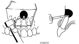
Along with the apicoectomy, the dentist usually performs a periapical curettage and may place a retrograde filling. After the patient has been draped and anesthetized, the dentist makes a surgical incision on the facial aspect of the alveolar ridge and a mucoperiosteal flap is elevated to expose the apex of the tooth to be treated. The assistant aids in retraction of the mucoperiosteal flap with the periosteal elevator and provides suction. This reveals the cortical bone of the alveolus covering the apex of the tooth. The dentist then uses a handpiece and surgical bur to remove the cortical plate covering the apex of the tooth. Once the root is exposed, the dentist uses the bur to remove the apex as shown in figure 7-21. The assistant irrigates with a saline solution and aspirates as needed. The dentist uses a curette to curettage the surrounding

Figure 7-21. - Removing apex of tooth.
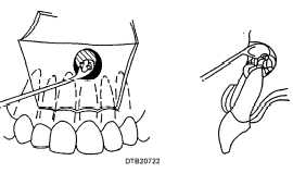
periapical tissue, thus removing infectious material from around the root tip. Figure 7-22 illustrates the curettage procedure. If access to the canal is obstructed from the occlusal or lingual aspect, debridement and filling can now be done from the apex.
If indicated, a retrograde filling may be performed to seal the apical end of the root canal by placing a

Figure 7-22. - Apical curettage procedure.
Continue Reading
