The entire dental team must agree to be consistent in charting methods. This is a slow process and much attention must be paid to details. Remember to check and double check each step. The team will decide which charting system they will use. The different branches of the service and civilian dentists all use different charting systems and abbreviations. The
Manual of the Medical Department, chapter 6, describes the Navy's charting system and abbreviations used to complete all dental information for the different forms used in forensic dentistry. Other dental abbreviations used for charting, such as the Computer Assisted Postmortem Identification, may be used and will be covered later in this chapter under Computer Support. The use of a fiberoptic light is invaluable in the examination process. The examiner begins by evaluating tooth #1 and associated radiographs. The second dentist on the examination team evaluates tooth #1 and confirms the findings of the first dentist. The recorder charts the findings of tooth #1 and all three members confirm the charting. Tooth #2 is examined and the process is repeated until all 32 teeth have been charted. The approach is redundant, but errors are corrected as they are made. Charting should be done in pen, not pencil. Findings to be recorded during the postmortem examination are as follows:
Dental restorations
Missing teeth
Prosthetic appliances
Pathology
Unique anatomy
Age estimate
References to possible gender and racial group
Teeth missing because of the trauma of the mishap should be specifically noted to avoid confusion over extracted or congenitally missing teeth. A prostho- dontist should be available to examine and describe dental prosthetic appliances. In some cases, the appliance may have been specifically marked for identification as shown in figure 10-15. It is wise to solicit from the victim's family study models or extra prosthetic appliances that may be available. Such evidence is important in providing antemortem data regarding ridge shape/size, rugae, and general oral anatomy. The antemortem dental record will be covered next.
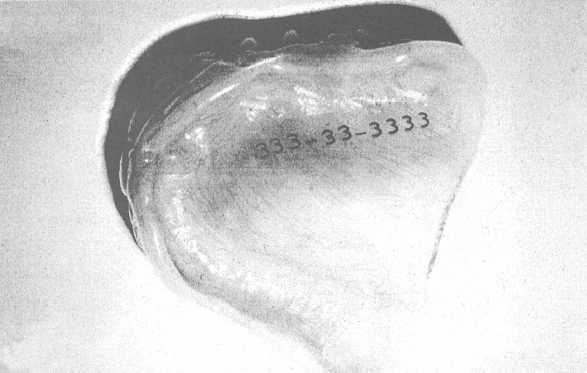
Figure 10-15. - Maxillary denture with SSN embedded in acrylic.
Continue Reading