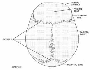
Figure 3-2. - Sutures of a skull.
Table 3-1. - Bones of the Cranium
| Single Bones | Paired Bones |
| Occipital | Parietal |
| Frontal | Temporal |
| Sphenoid | |
| Ethmoid |
the nasal cavity. In children, the frontal bone develops as two parts. They are usually fused together by age 5 or 6. The two frontal sinuses (air spaces in the bone) are located above each eye socket.
Parietal Bones
The two parietal bones are located behind the frontal bone. These bones form the greater part of the right and left sides and the roof of the skull. They each have four borders and are shaped like a curved plate.
Temporal Bones
The temporal bones form the sides and part of the base of the skull in the area of the ear. One temporal bone is located on each side of the head. It is readily recognized as “fan-shaped.” Each encloses the internal ear structures and have depressions called glenoid fossae that forms the articulation with the mandible.
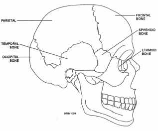
Figure 3-3. - Cranial bones.
The zygomatic process of the temporal bone projects out into the zygomatic bone of the face and forms the lateral part of the zygomatic arch. Both the glenoid fossae and zygomatic process can be seen in figure 3-4.
Occipital Bone
The occipital bone forms the back part of the skull and the base of the cranium. It joins with the parietal and temporal bones. In the center, underside (inferior) portion of the cranium, there is a large opening called the foramen magnum (fig. 3-5), through which nerve fibers from the brain pass and enter into the spinal cord.
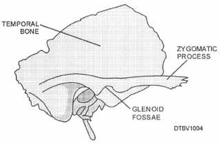
Figure 3-4. - Temporal bone.
Continue Reading