toward the inner angle of the eye where they drain down ducts into the nose.
MIDDLE LAYER.— The middle layer of the eye is called the choroid. It is a highly vascular, pigmented tissue that provides nourishment to the inner structures. Continuous with the choroid is the ciliary body, whose muscular structure attaches to the lens by means of suspensory ligaments and produces changes in the thickness of the lens. This permits the eye to focus to long-range or close-up vision.
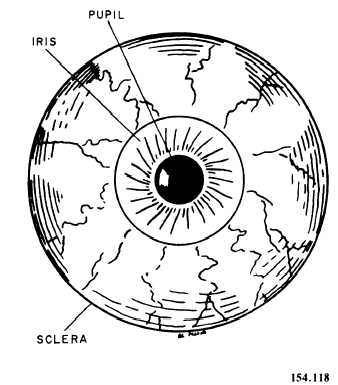
The iris is continuous with the ciliary body. It is a circular, pigmented muscular structure that gives color to the eye. The opening in the iris is called the pupil (fig 3-46). The amount of light entering the pupil is regulated through the constriction of radial/circular muscles in the iris. When strong light is flashed into the eye, the circular muscle fibers of the iris contract, reducing the size of the pupil. If the light is dim, the pupil dilates to allow as much of the light in as possible. The size and reaction of the pupils of the eyes are an important diagnostic tool.
The lens is a transparent, biconvex structure suspended directly behind the iris. It separates the interior eye into anterior and posterior cavities. The anterior cavity contains a watery solution called aqueous humor, which helps to give the cornea its curved shape. The optic globe posterior to the lens is filled with a jellylike substance called vitreous humor, which helps to maintain the shape of the eyeball and prevents misshaping by maintaining intraocular pressure.
INNER LAYER.— The inner layer of the eye is called the retina (fig. 3-47). It contains different layers of nerve cells, rods, and cones that are the receptors of the sense of vision. The retina is continuous with the optic nerve, which enters the back of the globe and carries visual impulses received by the rods and cones to the brain. The area where the optic nerve enters the eyeball contains no rods and cones and is called the blind spot. The rods respond to low intensities of light and are responsible for night vision. They are located in all areas of the retina, except in the small depression called the fovea centralis, where light entering the eye is focused, and which has the clearest vision. The cones require higher light intensities for stimulation and are most densely concentrated in the fovea centralis. The cones are responsible for daytime vision.

Figure 3-46.—Eye, anterior view.
Vision Process
Deflection or bending of light rays results when light passes through substances of varying densities in the eye (cornea, aqueous humor, crystalline lens, and vitreous humor) (fig. 3-48). The deflection is referred to as refraction. Accommodation is the process performed by the lens that increases or decreases its curvature to refract light rays into focus on the fovea.
The constriction of the pupil by the iris regulates the amount of light entering the eye. This process protects the retina from excessive stimulation and prevents a scattering of light rays that would produce blurred vision.
A movement of the globes toward the midline, which causes a viewed object to come into focus on corresponding points of the two retinas, is called convergence. This gives clear, three-dimensional vision.
The end receptors or nerve endings in the rods and cones that have been stimulated by light conduct impulses to the occipital lobes of the cerebrum, where they are interpreted into vision (fig. 3-48).

